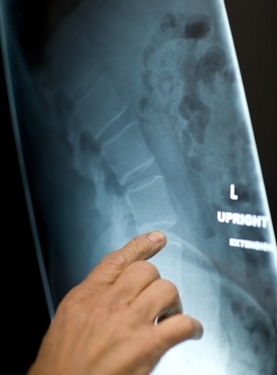
 Discography is currently used to determine whether the intervertebral disc is the source of pain in patients with back or neck pain.
During discography, contrast medium is injected into the disc and the patient's response to the injection is noted; provocation of pain that is similar to the patient's existing back or neck pain suggests that the disc might be the source of the pain. Computed tomography (CT) is usually performed after discography to assess anatomical changes in the disc and to demonstrate intradiscal clefts and radial tears.
The procedure:
Discography is usually performed under general anaesthetic. With CT guidance, needles are placed into the painful disc or discs, as well as a healthy disc adjacent to the diseased level, which acts as a control. The discs are then injected with a dye. You report the sensations and the CT scan records the anatomy of the disc disease. In addition to any general anaesthetic, the local nerves are blocked with local anaesthetic and adrenaline solution so as to reduce the amount of intra-operative bleeding and post-operative pain.
No instrumentation is implanted. You need only stay in hospital for 1 day which will most likely be at The Princess Grace Hospital.
If needed, we will arrange this all for you after your consultation at The Spine Surgery London.